Estudio de un caso de organoides derivados de pacientes (PDO) listos para usar en ensayos y adquisición de imágenes 3D de alto rendimiento para avanzar en el descubrimiento de fármacos
Introducción: el problema.
El coste medio de llevar un nuevo fármaco a la clínica es de aproximadamente 1000 millones de USD según un estudio realizado por la Facultad de Economía de Londres en marzo 2020 ( www.lse.ac.uk). Esto se debe en parte a los modelos poco representativos utilizados para la selección en las primeras etapas del descubrimiento de fármacos, lo que conduce a una alta tasa de fallos de los compuestos más adelante en la línea de producción de fármacos. Esto ha impulsado la búsqueda de modelos más centrados en el paciente para una selección temprana y precisa de los principales candidatos para un mayor desarrollo clínico.
Los Organoides derivados de pacientes (PDO o simplemente “organoides”) que se producen en tres dimensiones representan una solución a este problema. Son copias en miniatura del tejido de la biopsia humana normal o enfermado del que se derivan y representan completamente los tejidos 3D en el cuerpo humano. La evidencia muestra que cuando los pacientes y sus organoides derivados se tratan con los mismos fármacos, muestran una respuesta especular (Vlachogiannis et al., 2018). Esto muestra la prueba de principio de que los organoides se pueden utilizar para la selección de “bibliotecas” de posibles agentes terapéuticos. Es probable que los compuestos que tienen el efecto deseado en los organoides de los pacientes sean efectivos en el tratamiento de los propios pacientes.
¿Qué son los Organoides Derivados por el Paciente (PDO)?
- Derivado del tejido de la biopsia del paciente normal o dañado
- Cultivado en 3D en un gel proteico con alimentación líquida
- Formado a partir de varios tipos celulares (al igual que los órganos del cuerpo)
- Replicaciones de órganos en miniatura (p. ej., "miniintestinos")
- Totalmente representativo de la biología humana
- “Avatares” de patente para su uso en el descubrimiento de fármacos
- Cultivado en un bioprocesador para crear millones de copias normalizadas por lote
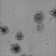
Imagen de campo claro de 3D, cáncer colorrectal, organoides derivados de pacientes (PDO)
Lo ideal es que, una vez que los fármacos se hayan probado correctamente y se hayan autorizado para su uso en la consulta, se diseñen tratamientos con fármacos individuales para cada paciente, en función de la respuesta de sus propios organoides. Por desgracia, actualmente se tarda varias semanas en extraer y expandir los organoides en el laboratorio. Los facultativos no querrían esperar ese tiempo antes de iniciar la terapia. La terapia personalizada puede ser posible en el futuro a medida que se refina y se desarrolle aún más la tecnología para derivar, cultivar y expandir los organoides.
Los organoides 3D son una tecnología relativamente nueva y los ensayos para hacer un uso completo de ellos todavía se están inventando y desarrollando. Los investigadores están más acostumbrados a trabajar con monocapas celulares 2D que son mucho menos complejas, pero que no representan con precisión la biología humana.
El reto: selección precisa de compuestos de las primeras pantallas, para un mayor desarrollo y pruebas clínicas.
El análisis de viabilidad es un método común para cuantificar la eficacia del compuesto (“eficacia”). Una técnica utilizada con frecuencia es un análisis basado en la bioluminiscencia que detecta la presencia de células activas de forma estática midiendo los niveles de ATP. Las imágenes de alto contenido también se pueden utilizar para determinar la viabilidad de las células mediante el recuento del número de células vivas o muertas (etiquetadas con tinciones como Calcein AM, un marcador común de células vivas) en una muestra tratada con fármacos en comparación con un control sin tratar (y posiblemente organoides derivados de tejido no relacionado con el cáncer). El efecto deseado de los fármacos utilizados en la terapia es matar tantas células cancerígenas como sea posible, dejando al mismo tiempo el tejido sano no afectado. El número de células que aún están vivas en la muestra no tratada será considerablemente mayor que en las células tratadas con el fármaco, especialmente cuando se utilizan altas concentraciones de la terapia. Por lo tanto, es importante cuantificar la diferencia entre las dos condiciones y determinar si los compuestos antineoplásicos como trametinib tienen eficacia. El mismo principio se puede utilizar para identificar nuevos agentes terapéuticos que funcionen eficazmente para matar las células cancerosas como parte de una pantalla de una biblioteca de compuestos.
No todos los fármacos actúan de la misma manera. Algunos fármacos actúan específicamente sobre un subconjunto objetivo de células o alteran las señales dentro de las células para evitar el nuevo crecimiento y la expansión de la enfermedad. No hay ningún efecto de eliminación de células a granel, por lo que las diferencias entre las células sanas y las células cancerígenas no se distinguen ni cuantifican fácilmente contando el número de células vivas restantes después del tratamiento. Se necesitan métodos alternativos.
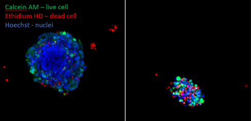
Los organoides del cáncer colorrectal se dejaron sin tratar (A) o expuestos hasta Trametinib (5mM) (B). Se tiñeron después de 5 días para mostrar las células vivas (verde, Calcein AM) y las células muertas (rojo, homodímero de etidio). Se puede ver claramente que quedan muy pocas células vivas después del tratamiento con trametinib.
La solución: para detectar los efectos discretos de los fármacos que actúan sobre objetivos específicos a través de imágenes 3D de alto rendimiento
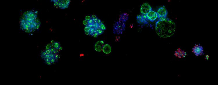
Los científicos han observado que el tratamiento de los organoides con fármacos puede provocar un cambio marcado de apariencia (morfología) que está relacionado con el efecto del fármaco en las células. Además de la viabilidad, se pueden añadir otros marcadores fluorescentes a la muestra para obtener información específica sobre las células y los orgánulos, como la estructura del citoesqueleto, las mitocondrias y la morfología general de los organoides. La captura de imágenes de organoides con imágenes de alto contenido y el uso de herramientas de análisis de datos para cuantificar los cambios, puede utilizarse potencialmente para indicar cuándo está teniendo un efecto un compuesto desconocido. Esto es especialmente relevante cuando un análisis de células vivas/muertas no puede detectar ninguna diferencia entre las células tratadas y las no tratadas. (Badder et al., 2020).

Ejemplo de cambios morfológicos capturados con el sistema microconfocal ImageXpress® de dispositivos moleculares . Los organoides del CCR se fijaron con una tinción fija con faroide (verde), Hoechst (azul) y homodímero de etidio (rojo). Tenga en cuenta los cambios en la mancha de faroide (verde) entre los grupos de control y de tratamiento
Para demostrar aún más el principio de que las imágenes de organoides en 3D se pueden utilizar en el descubrimiento de fármacos, Cellesce utilizó la automatización de última generación de dispositivos moleculares, la tecnología de imágenes y el análisis avanzado de los conjuntos de datos de imágenes en 3D para comparar las características físicas de los organoides de cáncer colorrectal sin tratar y tratados con fármacos. Se utilizaron herramientas de análisis de imágenes con ayuda de información artificial para analizar las imágenes CRC y se utilizaron varios descriptores fenotípicos para cuantificar los efectos del tratamiento. A continuación se muestra un ejemplo de una de las mediciones que se pueden realizar, es decir, el área total.

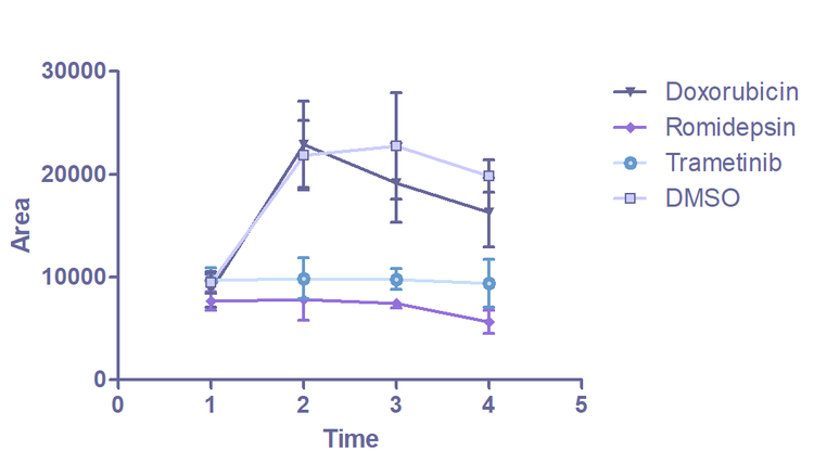
Imágenes de organoides capturados en el ImageXpress Confocal. Las imágenes de campo claro se utilizan para supervisar los cambios en la morfología de los organoides a lo largo del tiempo. El análisis se realiza en el software de análisis de imágenes IN Carta. Cada organoide identificado se superpone con una máscara de color. El gráfico (E) muestra el cambio en el área media de los organoides durante 5 días.
La mayor reducción en el área total se observa en los organoides tratados con los fármacos romidepsin y trametinib. En comparación con el control, el tratamiento con romidepsina y trametinib detuvo el crecimiento de los organoides. Este resultado es coherente con los datos del análisis de viabilidad, donde los organoides tratados con romidepsina y trametinib muestran un aumento significativo en las células muertas (como proporción del número total de células) en comparación con el control sin tratar. El análisis inicial de los datos de las imágenes ha confirmado la relación entre la morfología de los organoides y la respuesta al fármaco. Esto ha demostrado la prueba de principio de que esta técnica se puede utilizar en pruebas de detección de fármacos.
El uso de organoides y de imágenes en 3D tiene el potencial de revolución en la detección temprana en el descubrimiento de fármacos. Esto requiere loteos estandarizados y repetibles de grandes números de organoides. Dado que los organoides se cultivan manualmente, este requisito no se puede cumplir fácilmente. Cellesce ha abordado esta necesidad a través del desarrollo de un bioprocesamiento patentado. Esta nueva y exclusiva tecnología utiliza procesadores biológicos para la producción controlada de cantidades suficientes de organoides para la detección de alto rendimiento. Los dispositivos moleculares están abordando los requisitos adicionales para automatizar la manipulación de muestras mediante robótica y la optimización de ensayos mediante análisis de imágenes y datos.
El uso de organoides preparados para ensayos, junto con la automatización de ensayos de detección y cuantificaciones de alto rendimiento, facilitarán la selección precisa de productos terapéuticos de gran tamaño. Esto contribuirá en gran medida al proceso de identificación de tratamientos efectivos y aceleración de la línea de producción de detección de fármacos. La selección precisa de posibles compuestos terapéuticos en las primeras etapas del descubrimiento de los fármacos reducirá el desperdicio de recursos y el coste de desarrollo. Esto conducirá a un aumento en el número de fármacos en el mercado. Por lo tanto, los médicos serán capaces de elegir los tratamientos dirigidos que sean los más adecuados para cada paciente, con efectos secundarios adversos mínimos. Esto mejorará la calidad de vida y aumentará las tasas de supervivencia.
* fin *
Mejora del desarrollo de fármacos: El objetivo de los dispositivos moleculares y Cellesce es hacer avanzar el uso de los organoides
La reciente adquisición de Cellesce confirma el compromiso de los dispositivos moleculares con la inversión en tecnología de biología en 3D que transforma el proceso de detección de fármacos e impulsa el desarrollo de nuevos productos terapéuticos.
“Molecular Devices tiene la capacidad, la reputación, el alcance y los recursos para garantizar que la tecnología Cellesce se pueda desarrollar aún más y utilizar a todo su potencial”, dijo Vicky Marsh-Durban, Directora general de Cellesce. “Estamos encantados de llevar nuestra experiencia en el dominio y nuestra propiedad intelectual a los Dispositivos Moleculares, juntos para maximizar el impacto para los clientes en la revolución del descubrimiento de fármacos y desbloquear todo el potencial de la investigación en biología 3D relevante para el ser humano”.
Profundice en la conversación con Tanya Samazan, de Perspectivas de negocio de instrumentos, y la Presidenta de Dispositivos Moleculares, Susan Murphy, y el Director General de Cellesce, Vicky Marsh-Durban.

Descargue nuestro libro electrónico de Organoid
Si está listo para añadir otra dimensión a su investigación, la guía esencial de los organoides en el libro electrónico de descubrimiento de fármacos profundiza en la historia, el papel actual y el impacto futuro de los organoides en el descubrimiento de fármacos y cómo puede integrar correctamente los organoides en su investigación.