
Cómo desarrollar líneas celulares monoclonales para ingeniería de células madre
Garantizar la monoclonalidad es sin duda alguna crítico en el desarrollo de líneas celulares para la generación de tratamientos, como anticuerpos monoclonales o terapias a base de células. Para utilizar una línea celular para uso terapéutico, un investigador debe probar que las células de una colonia se derivan de una sola célula. El simple hecho de visualizar las colonias en los cavidades de la placa o utilizar un nivel de confianza en la monoclonalidad no constituye suficiente prueba de la misma si no hay una forma clara de realizar un seguimiento del crecimiento de las colonias a partir de una sola célula. Ahora, ¿qué más puede hacer para producir líneas celulares monoclonales y verificar su monoclonalidad?
Las soluciones CloneSelect ® de dispositivos moleculares presentan las tan necesarias tecnologías de flujo de trabajo para el desarrollo de líneas celulares monoclonales. Recientemente hemos hablado de los desafíos y oportunidades actuales para mejorar la ingeniería de la línea de células madre con Gargi Roy, científico del Departamento de Desarrollo Biofarmacéutico de AstraZeneca.
Anteriormente, Roy y su equipo de investigación utilizaron el ClonePix ® FL para producir anticuerpos monoclonales terapéuticos a partir de células de óvulo de hámster chino (CHO) [1]. Cuando cambiaron su enfoque a la ingeniería de las células madre, quedó claro que los dos flujos de trabajo tenían diferencias a pesar del rosca común de producción de anticuerpos monoclonales.
El equipo de terapia de células madre de AstraZeneca ha estado utilizando nuestros productos CloneSelect para la ingeniería de células madre y el desarrollo monoclonal. Además, Roy detalló cómo su equipo produce líneas celulares monoclonales con mayor precisión con la ayuda de las soluciones de detección de clones de Molecular Devices. Este artículo también cubre lo siguiente:
Descripción general de la ingeniería de las células madre
La ingeniería de las células madre implica la conversión de células somáticas, cualquier célula biológica excepto los semen y las células de óvulo, o las células embrionarias en células madre con capacidad para diferenciarse en todos los tipos de células. Este enfoque tiene como objetivo desarrollar varios modelos de células para el modelo de la enfermedad, la medicina personalizada, el descubrimiento de fármacos y los agentes terapéuticos basados en células madre, como los anticuerpos monoclonales.
Los dos enfoques principales de ingeniería de las células madre son los siguientes:
- Células madre pluripotentes inducidas (iPSC): Se convierten a partir de células somáticas mediante la introducción de factores de Transcripción.
- Células madre embrionarias (ESC): Derivado de células de masa interior embrionarias indiferenciadas.
Lo que ambos tipos de células madre tienen en común es que se pueden hacer palanca para producir anticuerpos monoclonales . Además, los anticuerpos monoclonales mediados por células madre poseen ciertas ventajas con respecto a los anticuerpos monoclonales recombinantes, como una mejor penetración en el área afectada y una mayor velocidad de cruce de la barrera hematoencefálica (BBB). [2].
Aislación de celda única con la impresora de celda única CloneSelect
La primera pregunta que uno podría hacer es, ¿qué hace que nuestras soluciones de flujo de trabajo CloneSelect destaquen en la realización de líneas celulares monoclonales y la posterior evaluación de la monoclonalidad? Imagínese poder depositar simultáneamente una sola célula en un depósito y garantizar la monoclonalidad con prueba visual en el momento de la clasificación.
La serie de impresoras de celda única CloneSelect garantiza un aislamiento de celda única sin problemas y facilita el crecimiento clonal máximo gracias a su suave dosificación de celdas, al tiempo que elimina la posibilidad de contaminación cruzada con otras líneas celulares. Su principio de trabajo radica en su tecnología microfluídica que encapsula suavemente las células individuales en gotas de líquido y las sembra en un depósito. Además, captura cinco imágenes por cada célula dispensada, lo que proporciona pruebas basadas en imágenes de aislamiento y deposición de una sola célula en el momento de la clasificación.
Para un sistema tan intrincado, la impresora de celda única CloneSelect es fácil de operar con nuestro software propietario. Una vez que su laboratorio complete una breve formación con nuestro equipo de personal de Científicos de aplicaciones de campo, se beneficiará inmediatamente de su diseño fácil de usar y fácil de usar para las celdas. El término “apto para celulares” se refiere a dos cosas:
- Ausencia de láseres de alta energía (la facilidad de uso de las células se logra a través de microfluídicas suaves alimentadas por gravedad)
- Volúmenes de dispensado diminutos (sin residuos y sin contaminación de muestra a muestra)
Tal vez lo más importante es que la adquisición de imágenes celulares de alta calidad por parte de la impresora de celda única CloneSelect se basa en sus criterios de exclusión, que se basan en la forma, el tamaño y la relación de eje de la célula. A través de los criterios de exclusión, puede clonar tanto las células parentales como las células modificadas con las propiedades deseadas.
Cómo crear líneas celulares monoclonales: El flujo de trabajo generalizado
Ya sea que trabaje con iPSC, ESC o muchos otros tipos de células, el flujo de trabajo resumido a continuación compone la base de la producción de la línea celular monoclonal:
- Impresión de celda única: Aísle una sola célula para iniciar una colonia
- Expansión y tipificación de la célula: Forme colonias y evalúe la monoclonalidad y asegúrese de que sus células expresan las proteínas de interés ejecutando ensayos genotípicos/fenotípicos
- Líneas celulares clonales: Diferenciación específica de tipo de célula
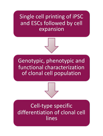
Un flujo de trabajo sencillo para comenzar con cualquier población de células, incluyendo su célula parental, y se utiliza para clonar algo o una célula diseñada para hacer que las células tengan cualquier propiedad deseada.
Retos en el aislamiento de una célula y el crecimiento clonal
Incluso con las tecnologías de aislamiento de célula única de última generación y los flujos de trabajo de monoclonalidad, todavía es posible encontrarse con algunos problemas.
Según Gargi Roy, el principal problema surgió para su equipo cuando querían imprimir células en múltiples placas y formar múltiples colonias con las mismas características . A medida que aumenta el número de repeticiones de aislamiento de célula única, a menudo se observa una caída constante en la eficiencia del crecimiento clonal. El declive es aún más pronunciado cuando se desea implementar condiciones sin componentes animales (ACF), que debe tener en cuenta si la investigación sin animales es crítica para su centro.
Las discrepancias en la eficiencia del crecimiento clonal externo son más importantes en los ESC que en los iPSC. La eficiencia cae a un solo dígito con ESC diseñados. La razón principal del declive es la caída en la viabilidad de la célula durante el enchapado, afirma Roy. También enfatiza la mejora del diseño experimental enriqueciendo el medio, especialmente en condiciones ACF.
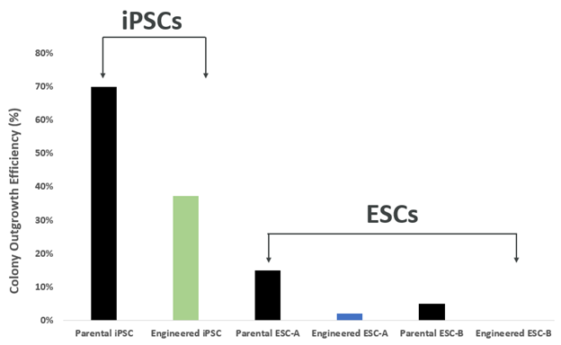
Las diferencias en las eficiencias de crecimiento de las colonias desde las células individuales entre los IPSC y los ESC ilustran discrepancias importantes en los ESC.
Incluso cuando hay suficiente crecimiento clonal, debe tener mucho cuidado para evitar la apoptosis. Una cosecha a tiempo de 8-10 días es fundamental. Ayudaría a alimentar su colonia a diario para evitar la inanición.
La agregación de las células es otro problema que obstaculiza la evaluación de la monoclonalidad. Debe utilizar el reactivo de disociación correcto para separar las células de la superficie del pozo y entre sí, lo que es un paso crítico para el análisis descendente.
Problemas de aseguramiento durante el desarrollo de la línea celular monoclonal
La verificación de la monoclonalidad es crucial para el desarrollo de las líneas celulares. Debe proporcionar pruebas detalladas de que su población de células se deriva de una sola célula para la presentación regulatoria. El equipo de Roy encontró que las ESC eran especialmente difíciles de supervisar debido a su superficie pegajosa y formas irregulares. Si se dispensaban dos ESC muy cercanos, es probable que se detecten como una célula individual. Además, si se enfrenta a una fecha límite ajustada, podría aplicar una dilución limitante para las estadísticas a fin de calcular la posibilidad de monoclonalidad.
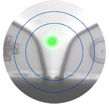
CloneSelect Single-Cell Printer ofrece técnicas de imagen avanzadas para probar la monoclonalidad en el punto de aislamiento de una célula . La impresora no solo puede distinguir fácilmente entre dos células muy cercanas durante la deposición de la célula, sino que también le permite rastrear la colonia desde el día de la cosecha hasta el día 0 para garantizar la monoclonalidad como se ve en la siguiente figura.
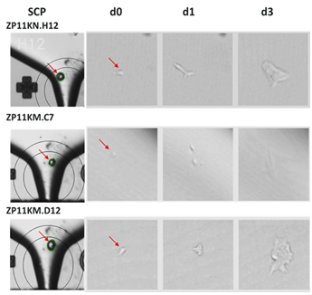
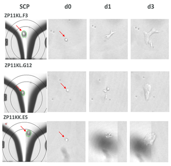
Confirmación de la monoclonalidad y captura de imágenes de célula única con la impresora de célula única CloneSelect
Caracterización clonal
El componente final del desarrollo de la línea celular es la tipificación. Una vez evaluada la monoclonalidad, se asegura de que esta se refleje en el genotipo y la pluripotencia. ¿Tienen todas las células de su colonia niveles uniformes de la funcionalidad deseada? Y lo que es más importante, ¿son estables desde el punto de vista genómico, lo que significa que pueden conservar y transferir material genético de una generación a la siguiente? Por último, ¿son pluripotentes (es decir, pueden diferenciarse en tres capas principales de microbios: ectodermo, endodermo y mesodermo)? Las respuestas solo se pueden obtener mediante la selección de clones de alto rendimiento.
Aquí es donde el generador de imágenes CloneSelect puede añadir valor a su investigación a través de una confianza adicional en la garantía de monoclonalidad. CloneSelect Imager contiene tecnología de campo claro sin etiquetas con imágenes de fluorescentes personalizables, junto con herramientas de análisis de datos avanzados, que incluyen mediciones de confluencia, curvas de crecimiento, generación de informes de monoclonalidad y miniaturas de placas para visualizar rápidamente el crecimiento de las colonias en cada pozo.
Con la alta resolución y la variedad de modos de imagen, incluso puede detectar los marcadores celulares específicos de las células de su clon . Esto le permite confirmar si sus líneas celulares monoclonales tienen la funcionalidad deseada.
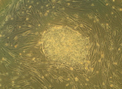
La siguiente ilustración muestra la diferenciación de las ESC monoclonales en tres capas de microbios diferentes, con los respectivos biomarcadores en cada capa.
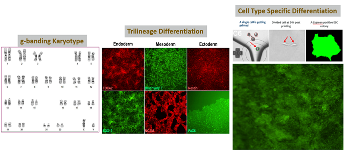
Células de alta calidad/estables utilizando nuestra impresora de célula única, lo que facilita la separación en tres capas de microbios: endodermo, mesodermo y ectodermo.
El alcance de la evaluación funcional del generador de imágenes CloneSelect continúa incluso después de comenzar la etapa de diferenciación de las células. A través de la generación de imágenes de fluorescente, puede monitorizar las células a medida que se diferencian para formar diferentes capas de tejido.
¿Cómo puedo Más información información sobre las líneas celulares monoclonales?
Desde la fase de siembra inicial hasta el crecimiento hacia afuera y la diferenciación de las células, el desarrollo de la línea celular monoclonal es más sencillo utilizando nuestro flujo de trabajo CloneSelect para la garantía monoclonal.
Puede seguir analizando la investigación del equipo de células madre de AstraZeneca para obtener más información sobre el flujo de trabajo de garantía de la monoclonalidad de la impresora de célula única CloneSelect y la impresora de cámara CloneSelect. En el reciente seminario web de Dispositivos Moleculares, Gargi Roy ilustra los problemas y las soluciones en la garantía de la monoclonalidad con detalles de su investigación que muestran cómo diseñar líneas celulares monoclonales y evaluar las células de su colonia.

Únase al seminario web a demanda, Aseguramiento de la monoclonalidad utilizando CloneSelect Single-Cell Printer y CloneSelect Imager en el flujo de trabajo de ingeniería de célula de tallo humana (hSC), con el Dr. Roy .
- Roy, Gargi, et al. "El tamizado secuencial por ClonePix FL y la tinción intracelular facilitan el aislamiento de líneas celulares productoras elevadas para la fabricación de anticuerpos monoclonales". Diario de métodos inmunológicos 451 ( 2017): 100-110.Roy, Gargi, et al. “El cribado secuencial mediante ClonePix FL y la tinción intracelular facilitan el aislamiento de líneas celulares de alto productor para la fabricación de anticuerpos monoclonales”. Diario de métodos inmunológicos 451 (2017): 100-110.
- Frank, Richard T et al. “Revisión concisa: las células madre como plataforma emergente para el tratamiento con anticuerpos contra el cáncer”. Células madre (Dayton, Ohio) vol. 28,11 ( 2010): 2084-7. doi:10.1002/stem.513.Frank, Richard T et al. “Revisión concisa: las células madre como plataforma emergente para el tratamiento con anticuerpos contra el cáncer”. Células madre (Dayton, Ohio) vol. 28,11 (2010): 2084-7. doi:10.1002/stem.513.